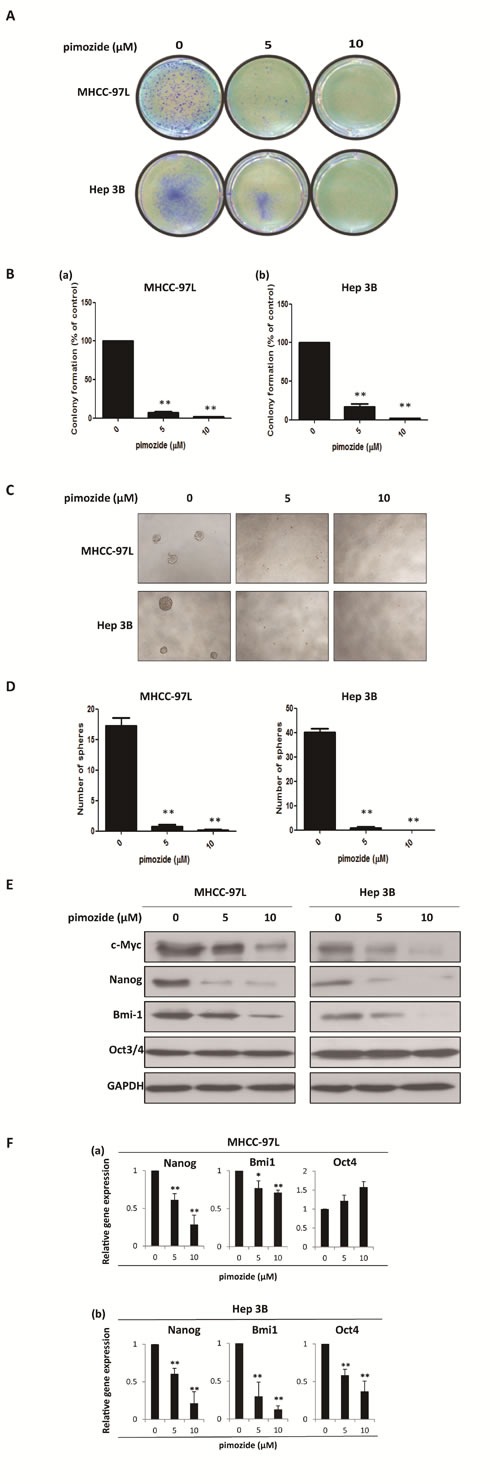
Figure 2. Pimozide inhibits the self-renewal capacity of HCC cells.

MHCC-97L and Hep 3B cells were treated with pimozide at the indicated concentrations, incubated for extra 10-14 day and then subjected to colony formation assay. Images were taken at a magnification of 100× A. The numbers of colonies were counted after staining with crystal violet and the histogram indicated the number of colonies. The results are from 3 independent transfection experiments (B). (C & D). Sphere formation assay of HCC cells treated with pimozide. The spheres were imaged under a light microscope (magnification, 100× ), and the statistical results are shown. E. Western blot analysis of the expression of self-renewal genes. Cell extracts were probed with antibodies against c-Myc, Bmi1, Nanog, Oct3/4 and GAPDH. F. MHCC-97L and Hep 3B cells were incubated with the indicated doses of pimozide for 48h before subjected to RT-PCR to detect the expression of the self-renewal genes Bmi1, Nanog and Oct4. *p < 0.05, **p < 0.01, compared with the control.
