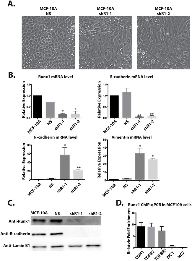
Figure 6. Depleting Runx1 in MCF10A cells promotes a mesenchymal-like phenotype.

A. MCF10A cells treated with shRunx1 show morphological changes toward an EMT- like state. B. Western blot analyses of lysates from MCF10A cells treated with shRunx1 show decreased protein expression of Runx1 and E-cadherin. C. RT-qPCR analyses of RNA from MCF10A cells treated with shRunx1 show decreased gene expression of E-cadherin and activation of mesenchymal marks of N-cadherin and Vimentin. Student's t test * p value <0.05, ** p value <0.01 for MCF10A shRunx1 cells compared to the MCF10A ns cells. Error bars represent the standard error of the mean (SEM) from three independent experiments. D. ChIP-qPCR confirmation of Runx1 occupancy at CDH1, TGFB2 and TGFBR1. ZNF188 (NC1) and ZNF333 (NC2) were used as the negative control as Runx1 are predicted not to bind these genes. Data obtained with antibodies against Runx1 are normalized to input control.
