
Figure 1. Capz inhibits the STAT3 signaling pathway by inhibiting constitutive JAK1/2 and Src activation.
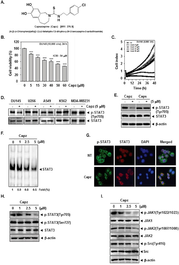
A. The chemical structure of Capsazepine (Capz). B. DU145 cells (1 × 10⁴ cells/well) were treated with the indicated concentrations of Capz for 24 h and cell viability was determined by MTT assay. C. The cell proliferation assay was performed using the Roche xCELLigence Real-Time Cell Analyzer (RTCA) DP instrument (Roche Diagnostics GmbH, Germany). DU145 cells (5 × 10³ cells/well) were seeded onto 16-well E-plates and continuously monitored using impedance technology. D. DU145, U266, A549, K562, and MDA-MB231 cells (1 × 10⁶ cells/well) were treated with Capz (5 μM) for 6 h. Whole-cell extracts were prepared and immunoblotted with antibodies for p-STAT3(Tyr705) and STAT3. E. DU145 cells (1 × 10⁶ cells/well) were treated with Capz or Caps (5 μM) for 6 h. Whole-cell extracts were prepared and immunoblotted with antibodies for p-STAT3(Tyr705) and STAT3. F. DU145 cells (1 × 10⁶ cells/well) were treated with the indicated concentrations of Capz for 6 h and nuclear STAT3 levels were measured using EMSA. G. Capz inhibited phosphorylation and translocation of STAT3 to the nucleus. DU145 cells (4 × 10⁴ cells/well) were incubated with or without 5 μM Capz for 6 h and intracellular p-STAT3 and STAT3 distributions were analyzed by immunocytochemistry. H. DU145 cells (1 × 10⁶ cells/well) were treated with 0, 1, 2.5, or 5 μM Capz for 6 h. Whole-cell extracts were prepared and immunoblotted with antibodies for p-STAT3(Tyr705) and p-STAT3(Ser727). The same blots were stripped and reprobed with STAT3 antibody to verify equal protein loading. I. Equal amounts of lysates were analyzed by Western blot using antibodies against p-JAK1(Tyr1022/1023), p-JAK2(Tyr1007/1008), and p-Src(Tyr416). The same blots were stripped and reprobed with JAK1, JAK2, and Src antibodies to verify equal protein loading.