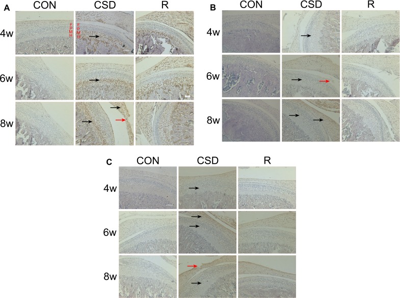
Figure 3. Expression of VEGF, Dll4 and p-ERK1/2 protein in the mandibular condylar cartilage of rat TMJs.
Representative images of immunohistochemistry staining for VEGF, Dll4 and p-ERK1/2 in the CON, CSD and recovery group (original magnification: ×200, scale bar = 100 μm). (A) Expression of VEGF protein in the rat TMJ mandibular condylar cartilage layers: fibrous layer (F), proliferating cell layer (P), mature cell layer (M), hypertrophic cell layer (H). VEGF-positive cells are indicated with black arrows and the damaged zone of the articular disc of TMJ is indicated with red arrows. (B) Expression of Dll4 protein in the rat TMJ mandibular condylar cartilage. Dll4-positive cells are indicated with black arrows and the damaged zone of the condylar cartilage is indicated with red arrows. (C) Expression of p-ERK1/2 protein in the rat TMJ mandibular condylar cartilage. Phosphorylated-ERK1/2-positive cells are indicated with black arrows and the damaged zone of the articular disc of TMJ is indicated with red arrows. (R: group).

