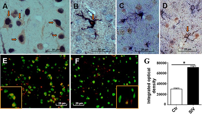
Figure 2. Menin expression was primarily observed in the nuclei of the frontal cortex neurons in SIV-infected macaques.

Menin expression (dark blue) is mainly observed in the nuclei of neurons (NeuN brown) in the frontal cortex of SIV-infected macaques A. Menin (dark blue) is also positive in activated microglial cytoplasm and processes, but not in microglial nuclei (Iba1, red) B. Menin (brown) is rarely stained in astrocytes of the cerebral cortex (GFAP, dark blue) C. but is positive in membranes and processes of white matter astrocytes (GFAP, dark blue) D. Representative double-labeled immunofluorescence images show NeuN (green) and menin (red) expression in the frontal cortex E–F. Menin expression is increased in SIV-infected macaques (E) compared with control macaques (F). Analysis of IOD of the double-positive area (yellow) shows significantly increased menin expression in neuronal nuclei of the SIV-infected macaques (#2, 4, 7, 8) compared with control macaques (#10–13) G. Data are expressed as mean ± SD, *P < 0.05. Original magnification: (A–D) 800×, (E–F) 400×. (A) From macaque #7; (B) from macaque #1; (C, D) from macaque #3. (E) from macaque #7; (F) from macaque #13. Arrows showing positive cells of double IHC staining.
