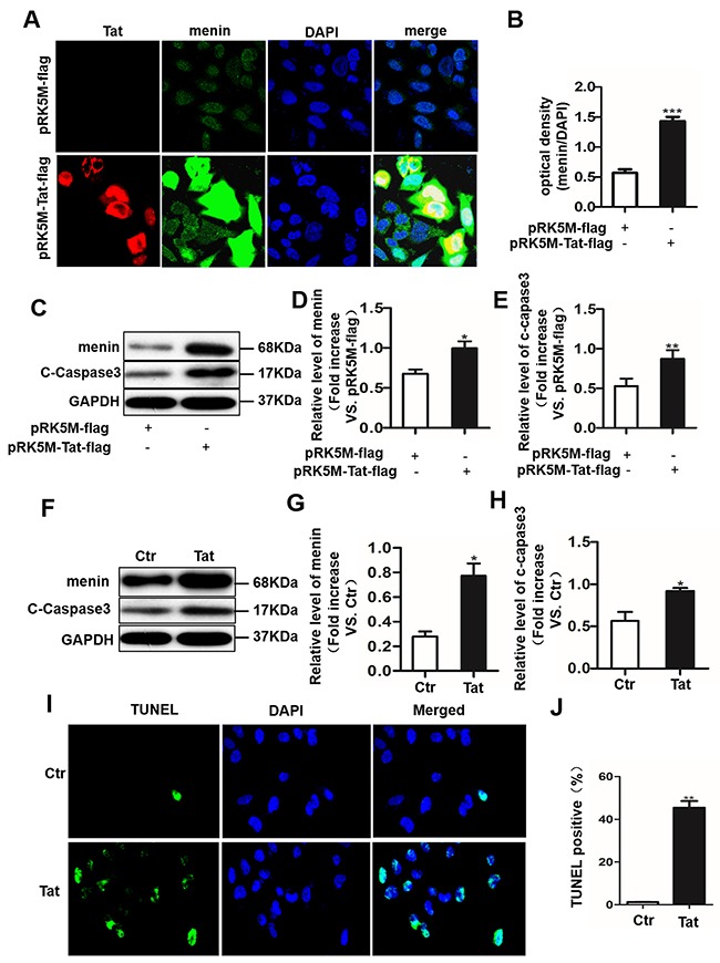
Figure 4. Tat induces cell apoptosis with an enhanced menin expression in SH-SY5Y cells and primary neurons.

SH-SY5Y cells were transfected with pRK5M-Tat-flag or pRK5M-flag for 24 hours and subjected to IHC staining. Menin (green) and Tat (red) co-localized in the nuclei of SH-SY5Y cells A. Optical density assay showed that menin expression is significantly increased in SH-SY5Y cells transfected with pRK5M-Tat-flag compared with the group transfected with pRK5M-flag B. Representative western blot were shown C. Optical density analysis of menin expression D. Optical density analysis of cleaved caspase3 expression E. Primary neurons were treated with or without Tat (100 ng/mL) for 48 h. Menin and cleaved-caspase 3 is significantly increased in Tat-treated neurons compared with controls F-H. TUNEL staining shows significantly increased apoptosis (green) in pRK5M-Tat-flag-transfected SH-SY5Y cells I-J. Data are plotted as mean ± SD (n = 3). *P < 0.05, **P < 0.01.
