
Figure 1. NICD downregulates ATXN1 expression.
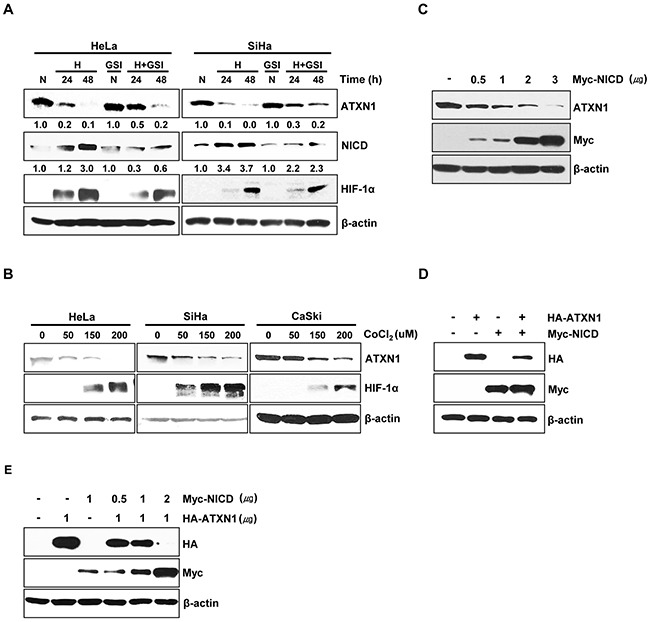
A. HeLa and SiHa cells cultured under normoxic or hypoxic conditions for 24 and 48 h in the absence or presence of the γ-secretase inhibitor GSI-DAPT were subjected to a western blotting analysis of ATXN1 expression. Densitometry results of ATXN1 are shown below each lane. ATXN1 expression was normalized to β-actin levels. Numbers indicate the intensity ratio relative to each control lane (1.0) B. HeLa, SiHa, and CaSki cells were treated with the indicated concentrations (μM) of CoCl2 for 24 h, and lysates were analyzed via western blotting with the indicated antibodies. C. HeLa cells transfected with Myc-NICD were subjected to western blotting analysis. D. Western blotting analysis of HEK293 cells co-transfected with HA-ATXN1 and Myc-NICD. E. Western blotting analysis of HEK293 cells co-transfected with HA-ATXN1 and the indicated amounts of Myc-NICD DNA. H (Hypoxia), N (Normoxia), GSI (γ-secretase inhibitor).