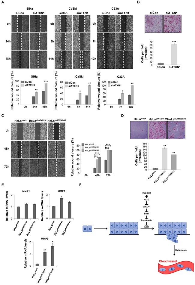
Figure 5. Inhibition of ATXN1 expression increases the migration and invasiveness of cervical cancer cell lines.

A. Upper panel: SiHa, CaSki, and C33A cells were transfected with siCon (sicontrol) or siATXN1 and subjected to a wound-healing assay. Lower panel: Quantification was performed by measuring the migration distances. *P<0.05, **P<0.01, ***P<0.001, t test. Scale bar: 100 μm. B. SiHa cells were transfected with siCon or siATXN1 and analyzed in a Matrigel invasion assay for 72 h. *P<0.05, **P<0.01, ***P<0.001, t test. Scale bar: 20 μm. C. The migration of HeLashATXN1-#1, HeLashATXN1-#2, and control cells was assayed in a wound-healing assay. *P<0.05, **P<0.01, ***P<0.001, t test. Scale bar: 100 μm. D. HeLashATXN1-#1, HeLashATXN1-#2, and control cells were analyzed in a Matrigel invasion assay for 72 h. *P<0.05, **P<0.01, ***P<0.001 vs. control group (one-way ANOVA). Scale bar: 20 μm. E. Real-time qRT–PCR analysis of MMP mRNAs in HeLashATXN1-#1, HeLashATXN1-#2, and control cells. All quantitative data are shown as the means and standard deviations of three independent experiments. *P<0.05, **P<0.01, ***P<0.001 vs. control group (one-way ANOVA). F. Proposed model for the role of ATXN1 in cervical cancer cell development. Hypoxia-induced upregulation of NICD expression induces reductions in ATXN1 and E-cadherin expression and an increase in Snail expression. Thus, ATXN1 expression decreases in the late stages of tumor development, thus inducing the EMT in cervical cancer cells.
