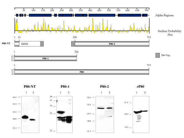
Figure 1.

Physical map and expression profile of recombinant P80 peptides. The 713 AA polypeptide chain of the P80 precursor protein is schematically represented by an alignment of proposed α helical and surface localized regions. The expressed regions in the different P80 variants are shown below in gray flanked by the amino acid numbers corresponding to the position within the P80 precursor. The striped boxes represent the fused poly His tags, colored in black or gray, respectively, depending on their presence or absence after purification of the recombinant protein. P80-NT was expressed as a fusion with dihydrofolate reductase (DHFR). In the Western-blot analysis shown below, lysates (lane 1) and purified P80 variants (lane 2) have been immunostained with an anti-His4-antibody (P80-NT), or the P80-specific monoclonal antibodies NB12 (P80-1) or LF8 (P80-2, rP80).
