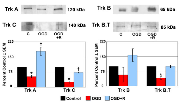
Figure 5.

Trk receptor protein expression in ONH astrocytes following exposure to and recovery from Oxygen-Glucose Deprivation. ONH astrocytes were exposed to OGD for 48 hours (OGD) or to 48 hours OGD followed by recovery for 24 hours (OGD+R). Cells exposed to growth media and 95% air/5% CO2 for 48 hours served as controls (C). Cell lysate was separated by SDS-PAGE and the density of unsaturated bands was measured for each blot. A representative blot for each Trk receptor is shown. Band density reported as mean percent of the control ± SEM (n = 3) is shown graphically beneath the blots. * indicates p < 0.016 for OGD compared to C and OGD+R; † indicates p < 0.016 for OGD+R compared to C (one way analysis of variance [ANOVA] followed by validation using Student-Newman-Keuls tests). Trk B.T; truncated Trk B.
