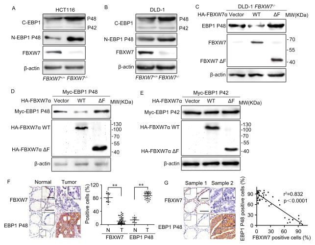
Figure 1. FBXW7 down-regulates EBP1 P48 protein level.
A and B. Western blotting on the whole cell lysates of HCT116 (A) and DLD1 (B) cells to confirm the protein levels of EBP1 isoforms with or without FBXW7 depletion. C. DLD1 FBXW7−/− cells transfected with HA-tagged FBXW7α or HA- tagged FBXW7ΔF. Western blotting confirmed that FBXW7 reduces endogenous EBP1 P48 level. D and E. HEK293T cells were co-transfected with HA-tagged FBXW7α or HA-tagged FBXW7ΔF and Myc-tagged EBP1 P48 (D) or Myc-tagged EBP1 P42 (E). Western blotting to assess the regulation of exogenous EBP1 isoforms by FBXW7. F. Immunohistochemistry staining of human colon tumor sections for EBP1 P48 and FBXW7 to show their expression differences in adjacent normal colon tissues and tumor tissues. Left: Representative images of FBXW7 and EBP1 P48 staining; Right: scatter diagram of relative expression of FBXW7 and EBP1 P48, Data as mean ± SD, Normal: adjacent normal colon tissues, Tumor: tumor tissue. N = 11 for normal tissues and 50 for colon tissues. G. EBP1 P48 expression was negatively correlated with FBXW7 expression in human colon cancer tissues. N = 50 for colon tissues. The scale bars in (F, G) represent 50μm and in the inserts of (F, G) represent 20μm. The results from (A–E) are repeated at least three times. ** P<0.01 based on Student t-test.

