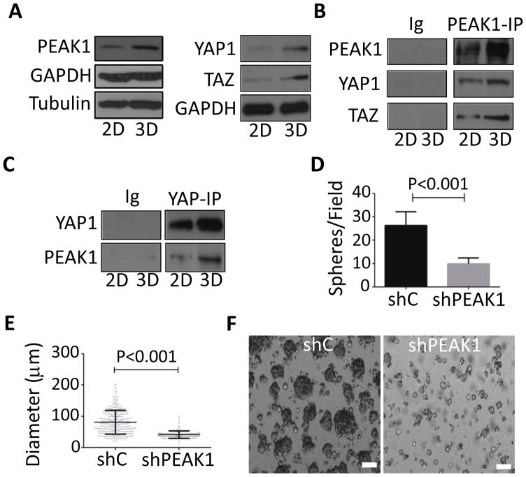
Figure 4.
3D sphere formation increases PEAK1-YAP1 complexes in PDAC cells. (A) 779E cells were cultured on plastic dishes in the presence of serum (2D) or placed in non-adherent culture dishes without serum to induce 3D sphere formation (3D) as described in Materials and Methods. Cells were then lysed and western blotted for the indicated proteins. (B and C) 779E cells treated as in A were lysed and analysed for PEAK1, YAP1, and TAZ complexes by co-immunoprecipitation (IP) and western blotting. Immunoglobulin coupled beads (Ig) served as a control. (D) The number of 3D spheres per microscopic field and (E) 3D sphere diameter was determined for 779E cells depleted of PEAK1 by shPEAK1 or treated with control shRNA (shC). (F) Representative phase-contrast photomicrographs of shC and shPEAK1 779E cells in 3D culture. Bar = 50μm.

