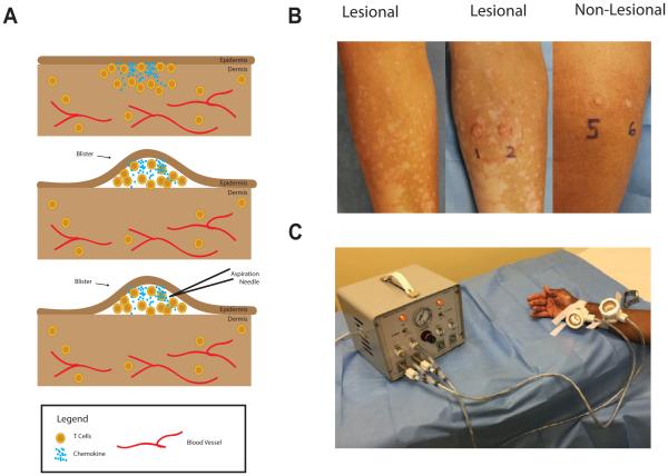
Figure 1. Modified Suction Blister Technique.
The process of suction blistering the skin (A): T cells cluster near the epidermis, where the chemokine concentration is highest (top panel). After the suction blistering, the epidermis forms the roof of the blister and is separated from the dermis. T cells and chemokine at the site are present in the blister fluid and can be aspirated by fine needle aspiration. Representative photos of active vitiligo before blistering (left panel), the same site after blistering (middle panel), and a non-lesional site from the same patient (right panel) (B). A study subject waits while the suction blisters form (C).

