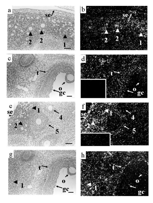
Figure 2.
Localization of expression of mRNA encoding TGF-β receptors in ovine ovaries. Panels a and b contain corresponding light field and dark field views of several small follicles from a 4 week old lamb following hybridization to the TGFβRI antisense RNA. Note specific hybridization in the oocytes of types 1/1a follicles (1) and type 2 follicles. Observe that some cells of the surface epithelium also express TGFβRI. Panels c and d contain corresponding light field and dark field views of a type 5 follicle from a 4 week old lamb following hybridization to the TGFβRI antisense RNA. Note the hybridization signal in the granulosa (gc), theca (t) and oocyte (o) of the type 5 follicle. Signal was also observed in many stromal cells. The inset in panel d contains a dark field view of the same area of the tissue hybridized to TGFβRI sense RNA. Observe the lack of specific concentration of silver grains over any cellular type. Panels e and f contain corresponding light field and dark field views of several small follicles from a 4 week old lamb following hybridization to the TGFβRII antisense RNA. Note the lack of specific hybridization in the type 1/1a and 2 follicles. Expression was observed in the theca of type 4 and 5 follicles however, note the lack of expression in the granulosa cells and oocytes of these follicles. Note also that some cells of the surface epithelium also express TGFβRII. The insert in panel f contains a dark field view of the same area of the tissue hybridized to TGFβRII sense RNA. Note the lack of specific concentration of silver grains over any cellular type. Panels g and h contain corresponding light field and dark field views of a type 5 follicle as well as several type 1/1a follicles from a 4 week old lamb ovary hybridized to the TGFβRII antisense RNA. Note that hybridization is limited to the theca (t) of the type 5 follicle and several stromal cells and is not observed in the granulosa cells (gc) or oocyte (o) of the type 5 follicle. In addition, signal is observed in the stroma around the type 1/1a follicles (1) but is not observed in the type 1/1a follicles. Scale bar equals approximately 100 μm for all panels.

