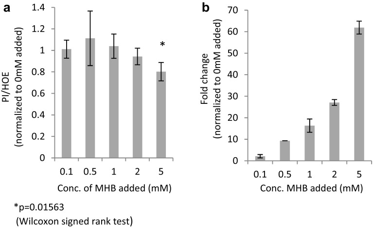
Fig. 4.
Cellular damage (n = 7) (a) measured as propidium iodide/hoechst33342 (PI/HOE) after reperfusion in the presence of 0.1, 0.5, 1, 2, and 5 mM (R)/(S)-methyl-2-hydroxybutanoate (MHB) during stabilization, simulated ischemia and reperfusion of cells and fold change (b) of intercellular concentration of α-hydroxybutyrate (AHB) (n = 2) measured semi-quantitatively in harvested cells (significance not calculated due to low number of included samples)

