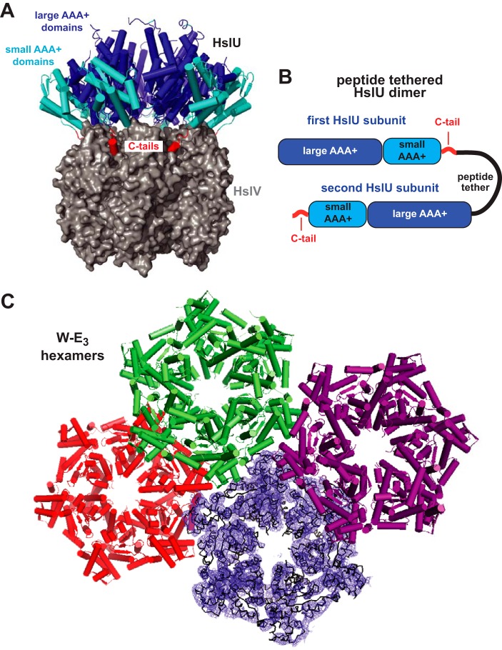
Figure 1.
HslUV structure. A, an HslU hexamer (secondary-structure representation) bound to an HslV dodecamer (surface representation; Protein Data Bank code 1G3I). The large and small AAA+ domains of HslU and its C-terminal tails are colored blue, cyan, and red, respectively. B, tandem HslU subunits connected by a genetically encoded peptide tether. C, three W-E3 hexamers in the asymmetric unit of structure 5TXV are shown in a secondary-structure representation; the fourth hexamer is shown in a ribbon representation with electron density from a composite omit map contoured at 1 σ.

