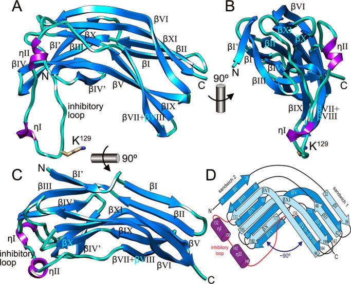
FIGURE 1.
Structure of the Kgp pro-domain. A, ribbon-type plot of Kgp-NPD showing the regular secondary-structure elements (310-helices in magenta, labeled ηI-ηII; β-strands as blue arrows, labeled βI′ + βI–βIV + βIV′ + βV–βXI; numbering based on the structure of RgpB-NPD; see Fig. 1 in Ref. 43). The inhibitory loop includes residue Lys129, which is depicted for its side chain and labeled. B and C, two orthogonal views of A. D, topology scheme of Kgp-NPD roughly in the same orientation as in C. Each regular secondary structure element is labeled and marked with its limiting residues.

