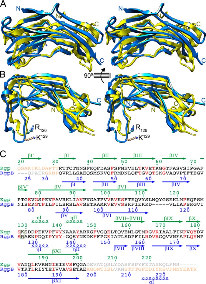
FIGURE 2.
Comparison of gingipain pro-domains. A, superposition in cross-eye stereo of Kgp-NPD (blue) and RgpB-NPD (yellow; PDB code 4IEF) (43) in the orientation of Fig. 1C. The respective N and C termini are labeled. B, orthogonal view of A. Here, the proven S1-intruding residue of RgpB (R126) and the putative one of Kgp (K129) are shown for their side chains and labeled. C, structure-based sequence alignment of the NPDs of RgpB and Kgp. Residues not defined in the respective structure are in light gray, structurally aligned residues are in black (when differing) or red (when identical), and residues defined but structurally non-equivalent in the two structures are in light salmon. The residue (potentially) intruding the S1 pocket of the respective CD is framed. Numbering and secondary structure elements (arrows for strands; kringles for helices) above the alignment in green correspond to Kgp; those in blue below the alignment correspond to RgpB.

