Figure 7.
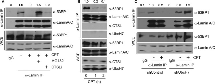
DNA damage reduced the interaction between 53BP1 and lamin A/C. A, A549 parental cells were treated with 500 nm CPT or vehicle in the presence or absence of 10 μm MG132 or 100 nm CTSLi for 8 h, and cell lysates were collected and immunoprecipitated (IP) with anti-lamin A/C antibodies, run on SDS-PAGE, and immunoblotted with anti-53BP1 or anti-lamin A/C antibodies. WCE were examined for protein expression. B, A549 parental cells were treated with 500 nm CPT for 0, 1, and 2 h, immunoprecipitated with anti-lamin A/C antibodies, and blotted with anti-53BP1, anti-lamin A/C, and anti-CTSL or anti-UbcH7 antibodies. Protein expression in WCE was also monitored. C, A549 cells stably depleted with control shRNA or shUbcH7 were treated with 500 nm CPT or vehicle for 8 h and processed as in B. The band intensities of the 53BP1 blots from both immunoprecipitate and WCE were quantified using Image J software, and the relative intensity of immunoprecipitated 53BP1 was normalized to that of 53BP1 proteins in WCE.

