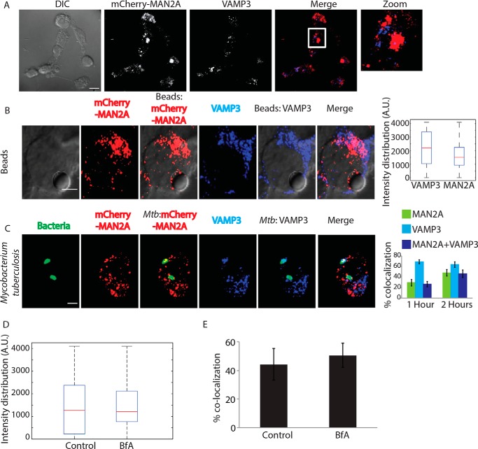
FIGURE 5.
Vesicles derived from Golgi apparatus are distinct from recycling endosome vesicles and are recruited independently. A, U937 cells expressing MAN2A-mCherry were fixed and stained with anti-VAMP3 antibody followed by Alexa Fluor 405-labeled secondary antibody (scale bar, 15 μm). White box in the merge panel identifies the area that was magnified for the zoom panel. DIC, differential interference contrast. B, mCherry-MAN2A (red)-expressing RAW264.7 macrophages were incubated with mouse serum-coated latex beads for 30 min or 1 h. At the respective time points, cells were immunostained with anti-VAMP-3 antibody followed by a secondary antibody tagged with Alexa Fluor 405 (blue). Presence of VAMP-3 or mannosidase-II at the bead surface was calculated using the 3D spot creation module in Imaris 7.2 software. The box plot at right shows data from more than 100 beads from two independent experiments (scale bar, 5 μm). C, mCherry-MAN2A (red)-expressing THP-1-derived macrophages were infected with PKH67-labeled H37Rv (green) for 1 and 2 h. At the respective time points, samples were fixed and stained with anti-VAMP-3 antibody followed by Alexa Fluor 405-tagged secondary antibody (blue). The images are representative of the 1-h time point. For the plots at right, % co-localization of H37Rv with mannosidase-II, VAMP-3, or both mannosidase-II and VAMP-3 was calculated using Imaris 7.2. The data represent an average of more than 150 bacteria from three different experiments (values ± S.D.; scale bar, 4 μm). D, U937 cells were incubated with serum-coated FITC-labeled latex beads in the presence or absence of BfA for 30 min. Samples were fixed and stained with anti-VAMP3 antibody followed by Alexa Fluor 405-labeled secondary antibody. Images were analyzed using 3D module in Imaris 7.2, and VAMP3 intensity distribution in the latex beads was calculated. E, U937 cells were incubated with GFP expressing E. coli in the presence or absence of BfA for 30 min. Samples were fixed and stained with anti-VAMP3 antibody followed by Alexa Fluor 405-labeled secondary antibody. Images were analyzed using 3D module in Imaris 7.2, and VAMP3 intensity distribution in the latex beads was calculated.

