
FIGURE 4.
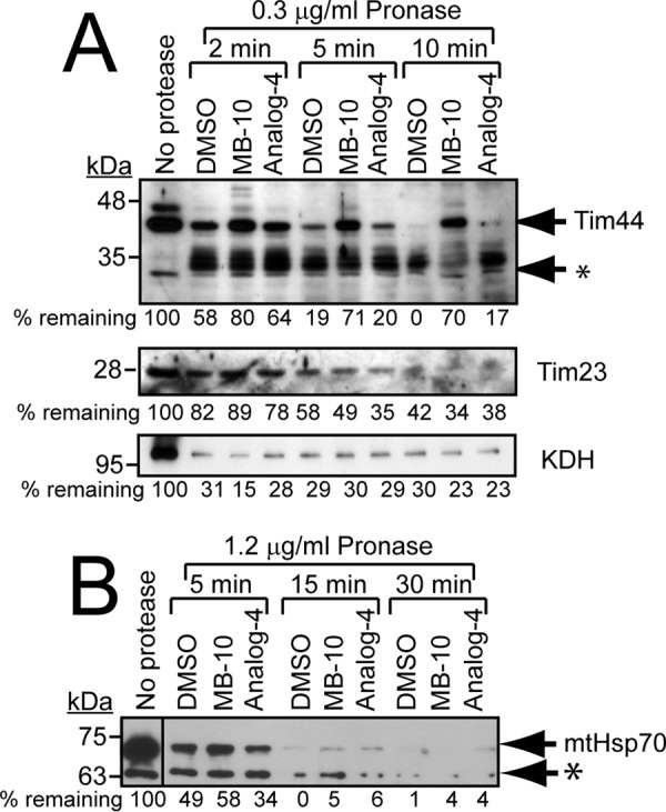
MB-10 targets Tim44. A, mitochondria were lysed in buffer containing 0.2% Triton X-100. The lysates were incubated with 1% DMSO, 100 μm MB-10, or 100 μm analog 4 for 15 min followed by treatment with 0.3 μg/ml Pronase at 25 °C. At the indicated time points, proteolysis was stopped by the addition of 0.2% SDS and incubation at 100 °C. Samples were analyzed by immunoblotting with antibodies against Tim44, Tim23, and α-ketoglutarate dehydrogenase (KDH). The asterisk indicates cleaved Tim44 products. A representative gel is shown; quantification of bands was performed with ImageJ software, and the percentage was calculated relative to the treatment with no protease. B, mitochondria were lysed in buffer containing 0.2% Triton X-100. The lysates were incubated with 1% DMSO, 100 μm MB-10, or 100 μm analog 4 for 15 min followed by treatment with 1.2 μg/ml Pronase at 25 °C. Samples were analyzed by immunoblotting with anti-mtHsp70 antibody. An asterisk mars a nonspecific band of lower molecular weight that was detected by the antibody.