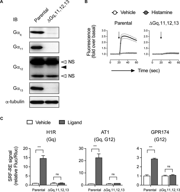
FIGURE 4.
Characterization of the CRISPR-generated quadruple Gαq/11/12/13-knock-out HEK 293 cells. A, Western blotting (IB) analysis of Gα expression. Note that there is a faint band for Gαq, which corresponds to an in-frame, loss-of-function mutant protein. Anti-Gα12 antibodies work poorly, and there were nonspecific bands (open arrowheads) near the faint endogenous Gα12 band (closed arrowhead). Similar results were obtained with two different commercially available antibodies (sc-409 (Santa Cruz Biotechnology) and 26026 (New East Biosciences)). B, Ca2+ influx assays using a FLIPR Calcium 5 kit to detect intracellular Ca2+ signal. Parental cells and the Gαq/11/12/13-knock-out line were transfected with a plasmid encoding the histamine H1 receptor and loaded with the Calcium 5 dye. The cells were treated with histamine or vehicle. C, SRF-RE reporter assays. We showed that the SRF-RE promoter assays (Promega) detect both Gαq and Gα12 responses. Parental cells and the Gαq/11/12/13-knock-out cells transiently expressing histamine H1 receptor, AT1R, or lysophosphatidylserine GPR174 (all in combination with the SRF-RE firefly luciferase plus CMV-driven Renilla luciferase) were treated with corresponding ligands. Fluc and Rluc signals were detected by dual measurement of both luciferases. Error bars, S.E.

