
FIGURE 6.
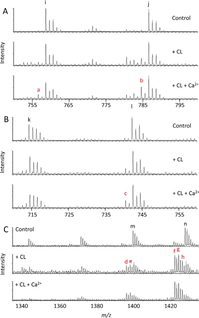
Mitochondrial tafazzin remodels cardiolipin by forward and reverse transacylations. Mitochondria were isolated from Sf9 cells expressing isoform A of Drosophila tafazzin. Isolated mitochondria (0.25 mg of protein) were incubated with 10 nmol of exogenous bovine heart CL with or without 15 mm CaCl2 for 90 min and analyzed by mass spectrometry. Exogenous CL triggered the formation of novel molecular species, including PC 16:1/18:2 (a; m/z = 756.6), PC 18:1/18:2 (b; m/z = 784.6), PE 18:1/18:2 (c; m/z = 740.5), CL (16:1)2-(18:2)2 (d; m/z = 1395.9), CL (16:1)2-18:1-18:2 (e; m/z = 1397.9), CL 16:1-(18:2)3 (f; m/z = 1421.9), CL 16:1-18:1-(18:2)2 (g; m/z = 1424.0), and CL 16:1-(18:1)2-18:2 (h; m/z = 1426.0). Native species of Sf9 mitochondria included PC 16:1/18:1 (i; m/z = 758.6), PC 18:1/18:1 (j; m/z = 786.6), PE 16:1/18:1 (k; m/z = 714.5), PE 18:1/18:1 (l; m/z = 742.5), CL (16:1)2-(18:1)2 (m; m/z = 1400.0), and CL 16:1-(18:1)3 (n; m/z = 1428.0). Addition of CaCl2 increased the concentration of several transacylation products. A, PC region (positive ion mode). B, PE region (negative ion mode). C, CL region (negative ion mode). Colored letters indicate novel molecular species.