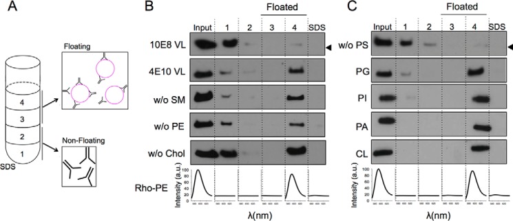
FIGURE 1.
Partitioning of the anti-MPER 4E10 Fab into membranes. A, 4E10 membrane partitioning detected in a sucrose gradient. Lipid vesicles incubated with the Fab were subjected to centrifugation. The sample was divided into four different fractions based on their different densities. An additional fraction employing SDS was collected, representing the material attached to the surface of the tube. The locations of the liposomes in the third and fourth fractions (i.e. floating fractions) were verified from the Rho-PE emission (bottom panels). The presence of Fab was probed by Western blotting. B, effect of the constituent lipids of the VL mixture on the process (lipid compositions are given in Table 1). 10E8 antibody was used as negative control (first row). C, effect of removing PS from the composition (first row) and recovery of membrane binding by its replacement with other anionic phospholipids: PG, PI, PA, or CL. The band corresponding to the Fabs comigrated in the gels with the 25-kDa molecular mass marker (indicated by the arrowheads, only in the first rows in B and C). SM, sphingomyelin; Chol, cholesterol.

