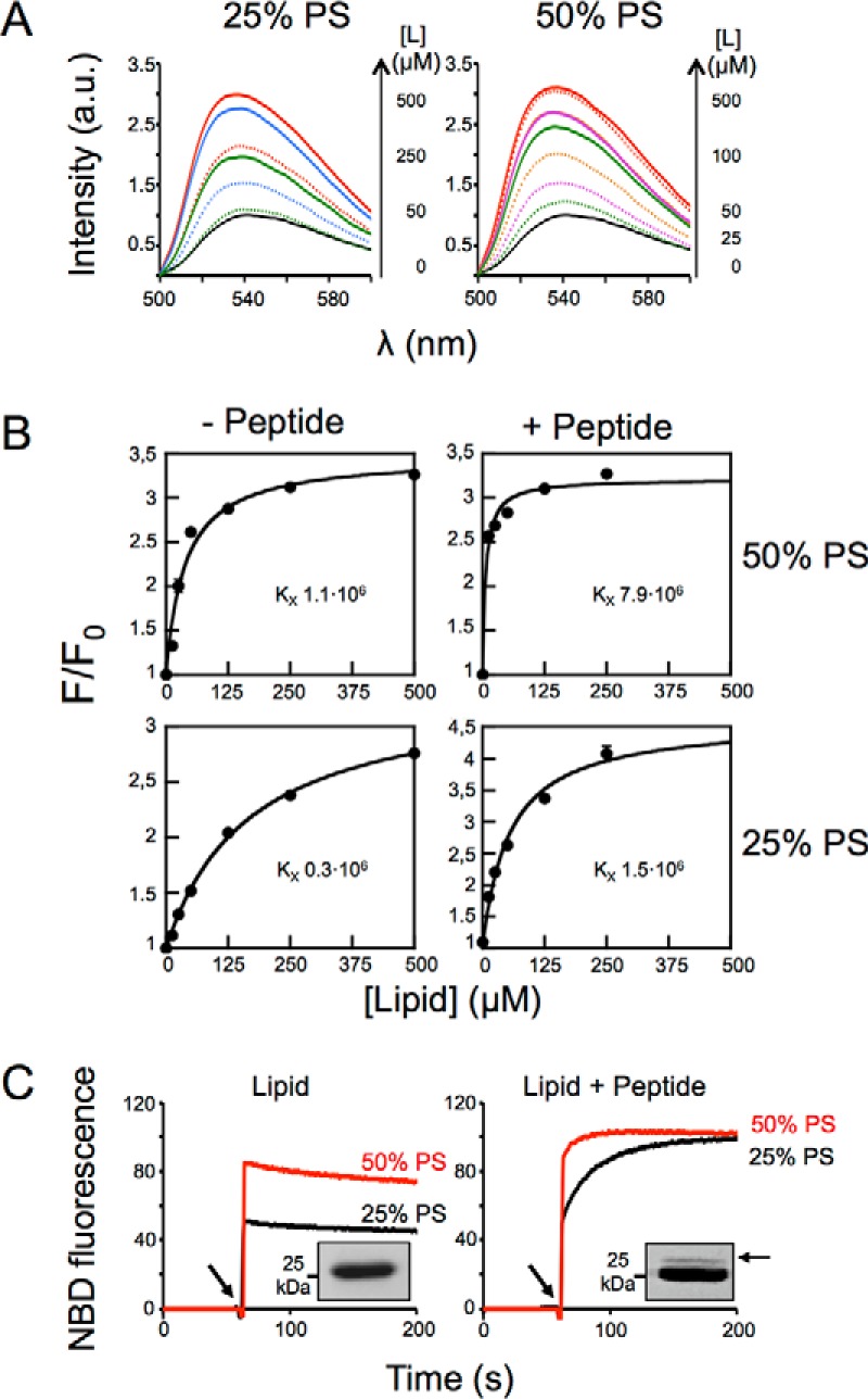
FIGURE 7.

Effect of electrostatic interactions on epitope recognition at the membrane surface. A, changes of NBD-4E10 fluorescence emission spectra in the presence of increasing concentrations of vesicles as indicated in the panel were measured in the absence (dotted lines) or in the presence of 1.7 μm of MPER(671–693) peptide inserted in the membrane (solid lines). B, effect of PS on Fab 4E10 partitioning into vesicles with and without MPER(671–693)peptide inserted in the membrane. NBD-Fab was titrated with PC:PS LUVs as indicated. Each data point corresponds to the average of three titrations (± S.D.) as the ones displayed in the previous panel. C, kinetics of incorporation of NBD-Fab into bare vesicles containing 25 or 50% of PS (left panel) or into the same vesicles decorated with 1.7 μm MPER(671–693) peptide (right panel). The arrow indicates NBD-Fab addition. Fab-peptide interaction was assayed by photo-cross-linking using Fab-pBPA and 25% PS vesicles. The presence of an adduct band confirmed Fab-peptide interaction in peptide-containing vesicles (insets). Lipid concentration was 100 μm. The results representative of two replicas are presented.
