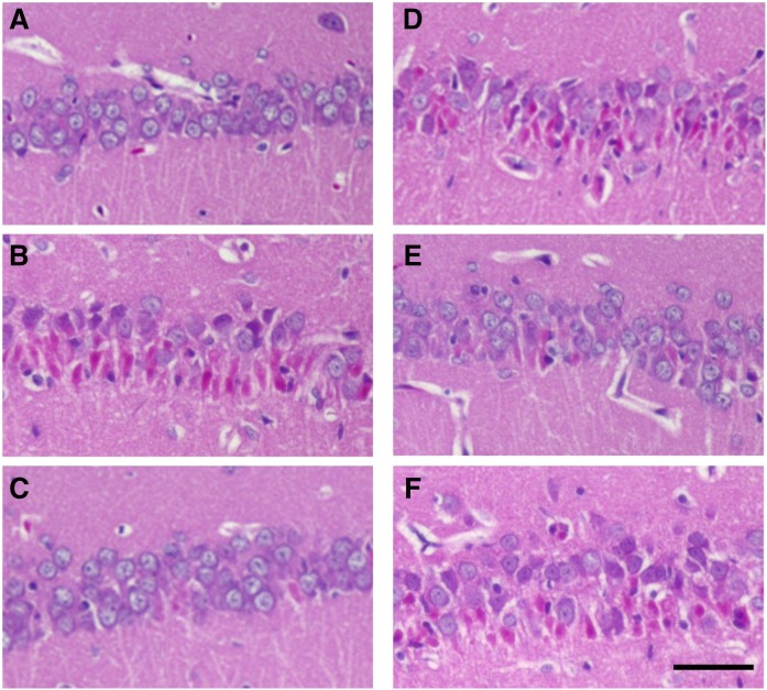
Fig. 1.
Histopathological sections (H and E staining) of the hippocampal CA1 region. Representative photomicrographs of coronal brain sections at the level of the fimbria demonstrate neuronal death of the hippocampal CA1 region to 30 days after the reagents administration are shown. H and E staining of control (A), KA (B), NS + KA (C), NS + KA + PGD2 (D), NS + KA + PGE2 (E), and NS + KA + PGF2α (F) groups is shown. Scale bar, 50 μm.

