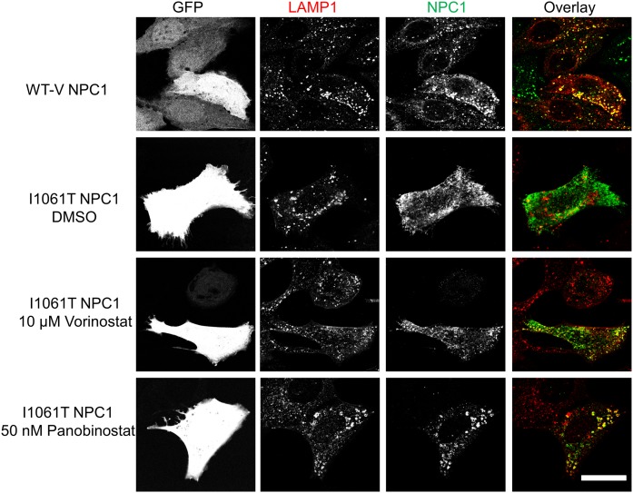
Fig. 6.
Vorinostat and panobinostat rescue localization of NPC1I1061T in U2OS-SRA-shNPC1 cells. WT-V NPC1 or NPC1I1061T was expressed in U2OS-SRA-shNPC1 cells by using a bicistronic vector also encoding eGFP. Starting 1 day later, wells transfected with NPC1I1061T were treated with 10 µM vorinostat, 50 nM panobinostat, or DMSO solvent control for 48 h. GFP serves as a marker of transfected cells. NPC1 protein was detected by immunofluorescence using anti-NPC1 rat monoclonal primary antibody and Cy5 goat anti-rat secondary antibody. Lysosomes were identified by using anti-LAMP1 rabbit polyclonal antibody and Alexa Fluor 546 goat anti-rabbit secondary antibody. The colocalizations of NPC1 protein and LE/Ly are shown in yellow. Scale bar, 10 µm.

