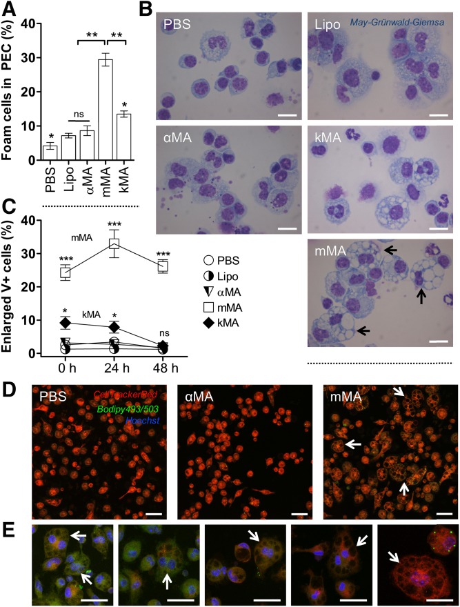
Fig. 2.
MAs of the methoxy oxygenation class promote the formation of multi-vacuolar foam cells. The proportion of vacuole-rich macrophages was determined by light microscope images of May-Grünwald-Giemsa-stained cells on cytospins and by laser-scanning-confocal microscopy (mean ± SEM). A: Percentage of foam cells in PEC fraction from the control (PBS and Lipo), αMA, mMA, and kMA treatments. (Shapiro-Wilk: W = 0.801, **P < 0.01; Kruskal-Wallis: H = 12.100, *P < 0.05, df = 4; n = 3 independent experiments; ns, not significant). B: Light microscope images of cytospins showing vacuolar foam cells in PEC fraction. Images were taken at 100× oil magnification. Arrows indicate MGCs. Scale bar: 10 μm. C: Induction of enlarged V+ cells is shown for control (PBS and Lipo) and the various MA-treated mouse peritoneal macrophages over time, as measured by laser-scanning-confocal microscopy (GLM: Wald chi-square = 753.924, ***P < 0.001, df = 14; n = 5 per time point; ns, not significant). D, E: Laser scanning confocal microscopy images showing enlarged V+ cells for PBS, αMA, and mMA treatments. Arrows indicate MGCs. Stacked images, 63× oil objective. Scale bar: 20 μm. E: Zoomed images from the mMA treatment in (D).

