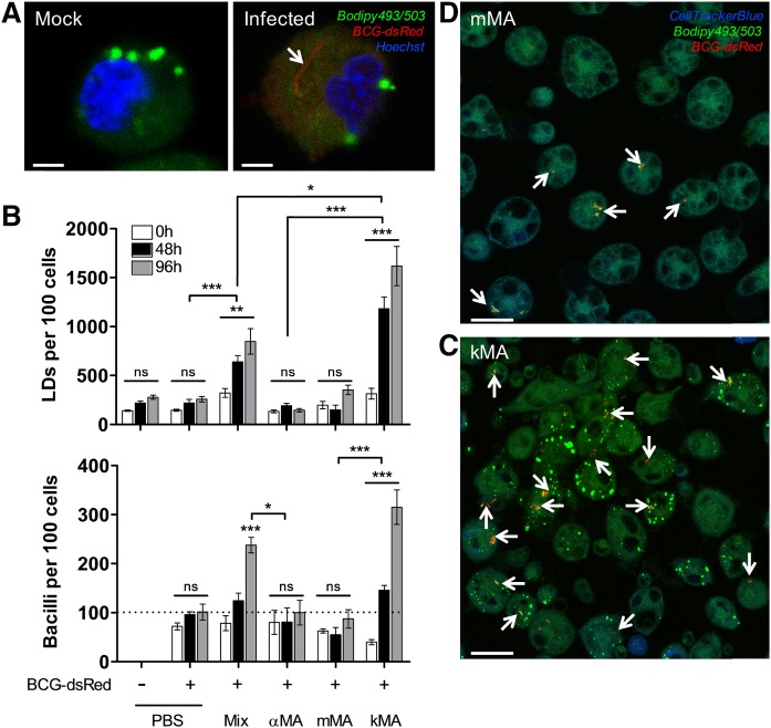
Fig. 4.
MA-induced LD accumulation in macrophages promotes BCG proliferation. Following in vivo treatment with MA, peritoneal macrophages were infected ex vivo with BCG-dsRed (MOI 1, broken line) and cultured until timed interval measurements were taken by laser-scanning-confocal microscopy (mean ± SEM). A: Laser-scanning-confocal microscopy images (zoomed) showing macrophages from mock or BCG infections. The presence of a BCG-dsRed bacillus can be clearly distinguished. Scale bar: 2.5 μm. B: Upper panel, LD accumulation in peritoneal macrophages over time (GLM: Wald chi-square = 636.496, P < 0.001, df = 17; n = 5 per time point). Lower panel, proliferation of BCG bacilli over time (GLM: Wald chi-square = 250.303, P < 0.001, df = 17; n = 5 per time point). On the x-axis, “Mix” denotes a natural purified MA extract consisting of ∼53% αMA, ∼38% mMA, and ∼9% kMA. C, D: Laser-scanning-confocal microscopy images depicting a clear difference in the presence of BCG-dsRed bacilli in peritoneal macrophages from mice treated with either kMA or mMA. Stacked images, 63× oil objective. Scale bar: 20 μm. Arrows indicate BCG-dsRed bacilli. Significant P values were ranked as *P < 0.05, **P < 0.01, and ***P < 0.001; ns, not significant.

