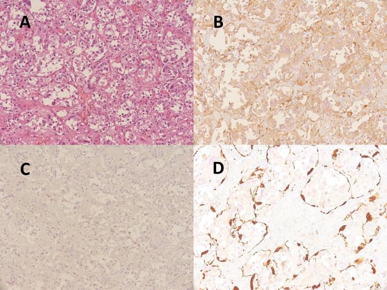
Figure 3.

Surgical specimen (A: haematoxylin-eosin staining, 200x) composed of two cellular types: chief cells, which have pale eosinophilic or clear cytoplasm with slightly to moderate atypical nuclei and are immunoreactive for synaptophysin (B: 200x) and negative for CKpool (C: 200x); and sustentacular cells, which have spindle-shaped nuclei with scanty cytoplasm and show immunoreactivity only for S100 (D: 400x), surrounding a nest of chief cells
