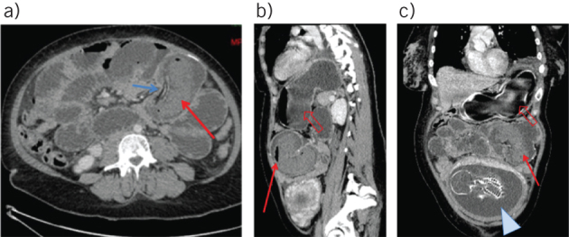
Figure 1.

Axial, sagittal and coronal contrast enhanced computed tomography of a 32-year-old woman with a history of Roux-en-Y gastric bypass: sausage-like mass appearance of intussusception (red arrow) with mesenteric vessels inside (blue arrow) (A); markedly dilated gastric remnant (open arrow), air–fluid levels in the intussuscipiens (solid arrow) (B); and fetus (arrowhead) (C)
