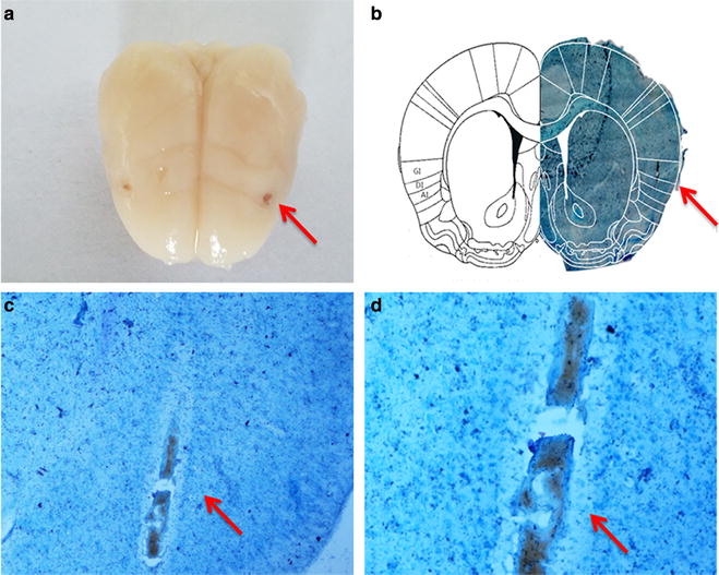
Fig. 4.

Nissl (a). The appearance of bilateral insular inserts (b). Comparison of position and mapping of the left lobes. Right a Nissl-stained frozen section of the rat brain (coronal cut, +1.2 mm relative to bregma [39]) was microinjected into the granular insular cortex as detailed in "Methods". Left a scheme of the corresponding contralateral hemisphere. GI granular insular cortex, DI dysgranular insular cortex. c ×200 target position of insular, d ×400 target position of insular
