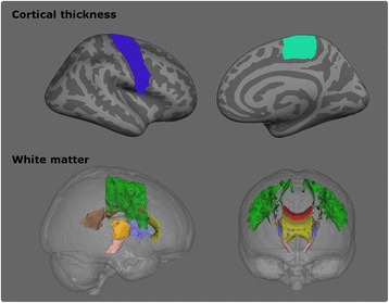
Fig. 3.

MRI features included in the regression analyses. Top: Cortical thickness. Blue: precentral gyrus, Light green: paracentral gyrus. Only the right hemisphere is displayed. Bottom: White matter. Green: superior corona radiata, grey: corona radiata, orange: posterior limb of the internal capsule, lilac: anterior limb of the internal capsule, rose: cerebral peduncle, yellow: genu of corpus callosum, red: body of the corpus callosum, brown: splenium of the corpus callosum
