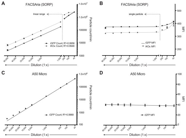
Fig 3.
Detection of single HIV-1 particles on the FACSAria II and A50 Micro. (A) Particle count/min of iGFP and iKOκ viruses by dilution factor on the FACSAria II. R2=0.9956 and 0.9948, respectively, p<0.0001 for both. (B) Mean fluorescent intensity (MFI) of iGFP and iKOκ viruses by dilution factor on the FACSAria II. (C) Particle count/min of iGFP viruses by dilution factor on the A50 Micro. R2=0.9968, p<0.0001. A lack of a laser with an appropriate excitation wavelength makes detection of iKOκ viruses impossible on the A50 machine used in this study. (D) Mean fluorescent intensity (MFI) of iGFP viruses by dilution factor on the A50 micro.

