Figure 4.
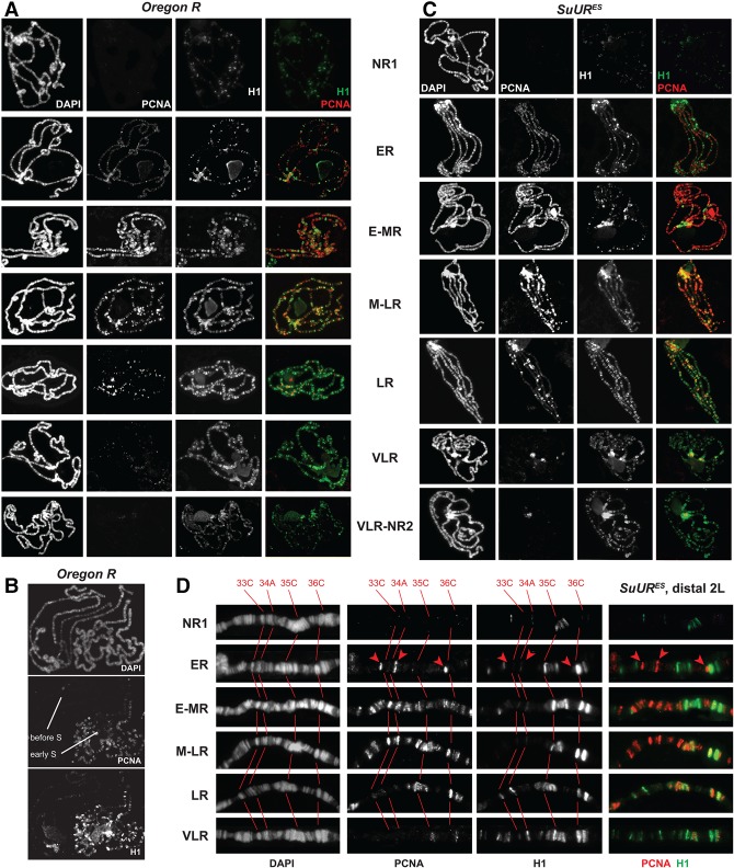
H1 distribution in polytene chromosomes is altered dynamically during endo-S phase. (A) Stage-dependent distribution of H1 during endo-S phase in wild-type polytene chromosomes. H1 (green) and PCNA (red) genome-wide distribution patterns were examined by IF staining of polytene chromosomes. The stages of endo-S phase were established based on PCNA distribution in the wild type according to Kolesnikova et al. (2013). H1 and PCNA exhibit mostly mutually exclusive patterns throughout the endocycle. DAPI staining shows the overall chromosome morphology. (NR1) No replication (before the onset of the endo-S phase); (ER) early replication (early endo-S phase); (E-MR) early to mid-replication; (M-LR) mid- to late replication; (LR) late replication; (VLR) very late replication; (NR2) no replication (after the completion of the endo-S phase). (B) Differential loading of H1 in polytene chromosomes before and at the onset of endo-S phase. Polytene chromosomes were stained and analyzed as in A. Two adjacent polytene spreads corresponding to “before” and early endo-S phase exhibit a dramatic difference in the level of H1 loading into chromosomes. (C) Stage-dependent distribution of H1 during endo-S phase in SuUR mutant polytene chromosomes. Polytene chromosomes were prepared from homozygous SuURES mutant larvae and analyzed as in A. (D) Stage-dependent distribution of H1 during endo-S phase in SuUR mutant distal polytene chromosome arm 2L. Detailed view of fragments of polytene chromosomes from (or similar to) those presented in C. DAPI staining shows the overall chromosome morphology and was used for an alignment of cytological positions. Red numbers at the top and corresponding cytological bands in grayscale panels are connected with red lines. (Red arrowheads) Early replicating interbands.

