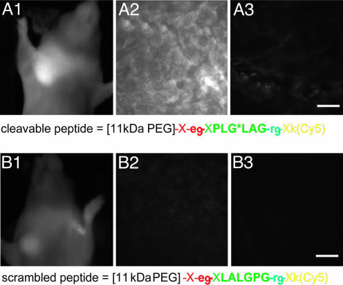
Fig. 4.
HT-1080 tumor xenografts visualized with activatable CPPs. HT-1080 tumors were implanted into the mammary fat pad of nude mice and allowed to grow until they reached 5-7 mm in diameter. (A1) A live anesthetized animal imaged 50 min after injection with 6 nmol of cleavable peptide. (A2 and A3) Tumor and muscle histology from a different animal killed 30 min after injection. (B1-B3) A similar experiment with the scrambled peptide. (Scale bars, 30 μm.)

