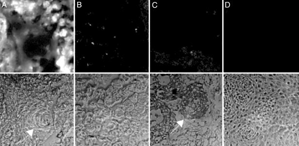
Fig. 5.
ACPP staining of surgically resected human squamous cell carcinoma tissue. Fresh tumor tissue was sliced in 1-mm slices and incubated in 1 μM cleavable (A) or uncleavable (B) peptide for 15 min, washed, and frozen. Sections (5 μm) were taken for fluorescence microscopy by using a 10× objective, and tissue type was verified by hematoxylin/eosin stain. (A) The arrow indicates a differentiated keratin pearl. As a control, histologically normal tissue from the same patient was treated similarly with MMP-2 cleavable peptide (C) or scrambled peptide (D). (C) The arrow indicates a ring of invading tumor cells.

