Fig. 2.
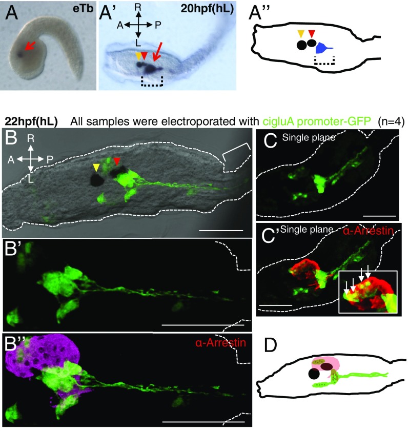
CiGluA is expressed in only a subset of neurons in the CNS. (A and A’) ciGluA expression analyzed using in situ hybridization. eTb, early tailbud stage, about 8.45 hpf (n = 26); hL, hatched larva at about 20 hpf (n = 31). The red arrow in A and the dotted brackets in A’ and A’’ indicate cigluA mRNA expression, the yellow arrowheads in A’ and A’’ indicate the otolith, and the red arrowheads in A’ and A’’ indicate the ocellus. (A’’) A schematic diagram illustrating the spatial relationship between cigluA mRNA-expressing cells and the otolith and ocellus in hL. (B–C’) Hatched larvae were electroporated with cigluA-GFP. (B) 3D reconstruction images showing merged DIC and GFP staining. The yellow arrowhead indicates the otolith, and the red arrowhead indicates the ocellus. The bracket indicates part of the tail. (B’) Higher magnification image of the GFP staining pattern in B. (B’’) Images costained for GFP and arrestin (a mature photoreceptor marker). (C) An independent sample expressing GFP and (C’) costained for arrestin. The white box in C’ is a higher magnification image of the photoreceptor region with arrows indicating cells that coexpress GFP and arrestin. (D) Schematic illustrating the spatial relationship between arrestin-positive photoreceptor cells (pink) and ciGluA-promoter–induced GFP-positive cells (green) in hL. The black circle and ellipse represent the otolith and the ocellus, and the dark green dots within the light green areas indicate cell nuclei. (Scale bars, 50 μm.)

