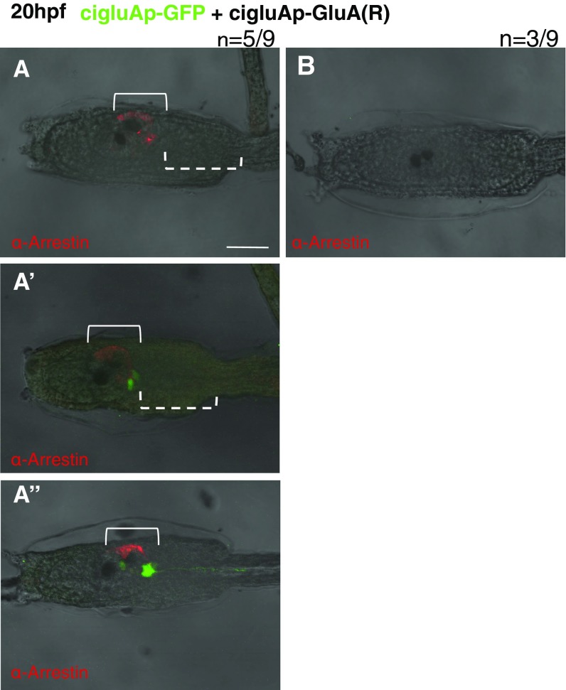
Fig. S6.
Various phenotypes seen in R-type GluA electroporated larvae (related to Fig. 4). (A–A’’) Representative images of larvae with partial loss of arrestin-positive photoreceptors. The solid brackets in A–A’’ show that R-type ciGluA-electroporated larvae have photoreceptor cells with partial loss of arrestin-positive cells. The dotted brackets show the region where the GFP signal disappeared (A) and the GFP-positive cells without their axons (A’). (Scale bar, 50 μm.) (B) Representative image of an R-type GluA-electroporated larva lacking expression of GFP and arrestin.

