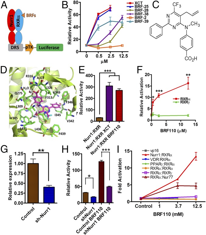
Fig. 1.
Discovery of BRF110, specificity for Nurr1:RXRα heterodimers, and i.p. administration in mice. (A) Schematic representation of the screening transactivation assay. (B) Typical results. (C) Chemical structure of BRF110. (D) Schematic representation of BRF110 interactions within the RXRα (PDB ID code 1MV9)-binding pocket. BRF110 (magenta) docked onto RXRα (green; PDB ID code 1MV9). Hydrogen bonds and amino acids are indicated. (E) Comparison of DR5-Luc induction by BRF110 (1 μM) and XCT (1 μM) via activation of Nurr1:RXRα heterodimers. (F) Cellular assay indicating that BRF110 activates Nurr1:RXRα heterodimers but not Nurr1:RXRγ heterodimers. (G) Retroviral-mediated knockdown of endogenous Nurr1 in SHSY5Y cells as measured by qPCR. (H) Activation by BRF110 of DR5-luciferase reporter plasmid introduced into SHSY-5Y cells infected with either a control retrovirus or the retrovirus carrying shNurr1 sequences. (I) Gal4-DNA–binding domain and Nurr1, Nur77, VDR, RXRγ, PPARγ, and RXRα ligand-binding domain fusions cotransfected with RXRα:vp16 fusions activated by BRF110 at the indicated concentrations.

