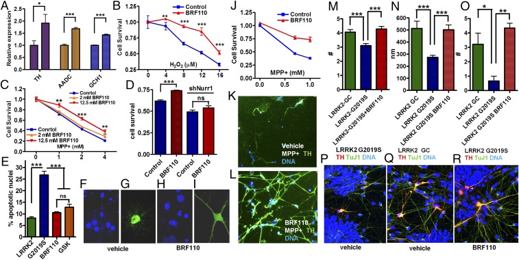
Fig. 2.
Induction of DA biosynthesis gene expression and of neuroprotection in cell lines and in patient iPSC-derived DAergic neurons. (A) Expression levels of the three DA biosynthesis genes (TH, AADC, and GCH1) mediated by the activation of Nurr1:RXRα by BRF110 (12.5 μM), as assessed by qPCR. n = 8. (B) SHSY-5Y cell viability increases with BRF110 treatment, as measured by the 3-(4,5-dimethylthiazol-2-yl)-2,5-diphenyltetrazolium bromide (MTT) assay, after exposure to varying concentrations (0–16 μM) of H2O2. (C) BRF110 concentration-dependent SHSY-5Y cell viability, as measured by the MTT assay, after exposure to varying concentrations (0–4 mM) of MPP+. (D) BRF110-mediated SHSY-5Y cell viability against MPP+ (2 mM) is dependent on Nurr1 levels, as determined by retroviral knockdown of Nurr1. (E) BRF110 treatment of primary rat cortical neurons reduces apoptotic death induced by cotransfecting LRRK2-G2019S or control WT LRRK2 cDNAs with CMV GFP, as measured by DAPI staining. (F–I) Representative DAPI (F and H) and GFP (G and I) confocal images. (J) Survival of human iPSC-derived DAergic neurons exposed to MPP+ (0.5 and 1.0 mM for 24 h) and receiving vehicle or BRF110. (K and L) BRF110 treatment preserves DAergic neuron projections, as determined by TH immunofluorescence of surviving iPSC-derived DAergic neurons after exposure to MPP+. (M–R) PD patient iPSC-derived DAergic neurons with the LRRK2-G2019S mutation or corrected mutation (GC). BRF110 treatment rescues neurite number (M), neurite length (N), and neurite branching (O) phenotypes. (P–R) Representative images stained with TuJ, TH, and DAPI.

