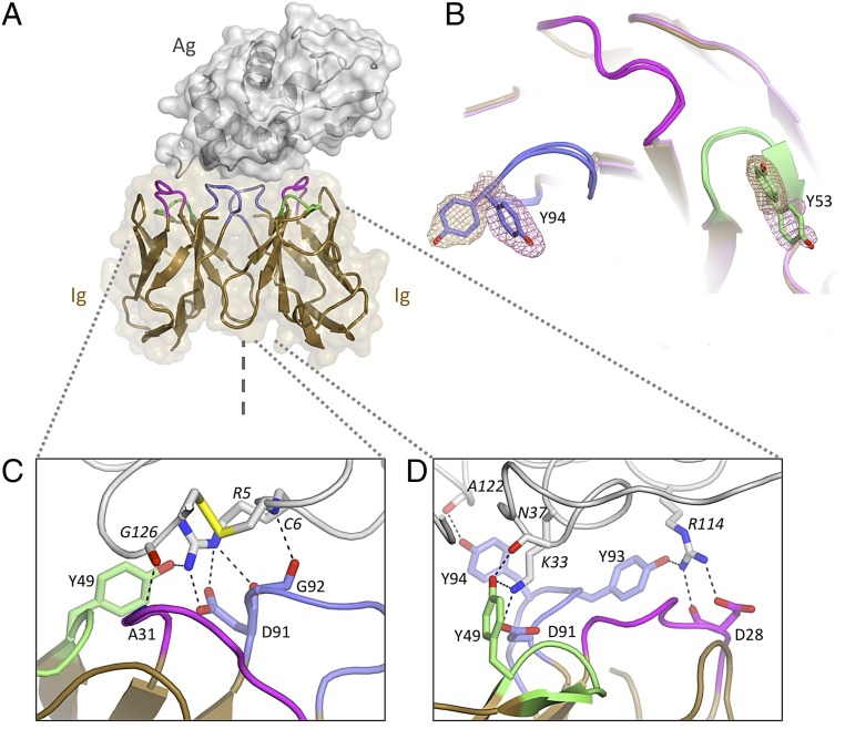
Fig. 2.
Structure of the Ig5 homodimeric antigen receptor in complex with antigen: Evidence for conformational diversity. (A) Homodimer of Ig domains (tan cartoon with CDR1, CDR2, and CDR3 loops colored in purple, green, and blue, respectively) bound to a single HEL molecule (gray cartoon and surface). The symmetry axis is indicated (dashed line). (B) Antigen-binding site (superposed Ig protomers, chains C/D): Multiple side-chain conformations are observed at positions Y53 and Y94. Electron density (mesh) is contoured at 1 SD above average (sigma-A weighted 2mFo-DFc map). (C) Antigen-binding site 1 (left-hand side). (D) Antigen-binding site 2 (right-hand side). Hydrogen bonds are highlighted; distinct contact networks are observed for each Ig subunit.

