Fig. 3.
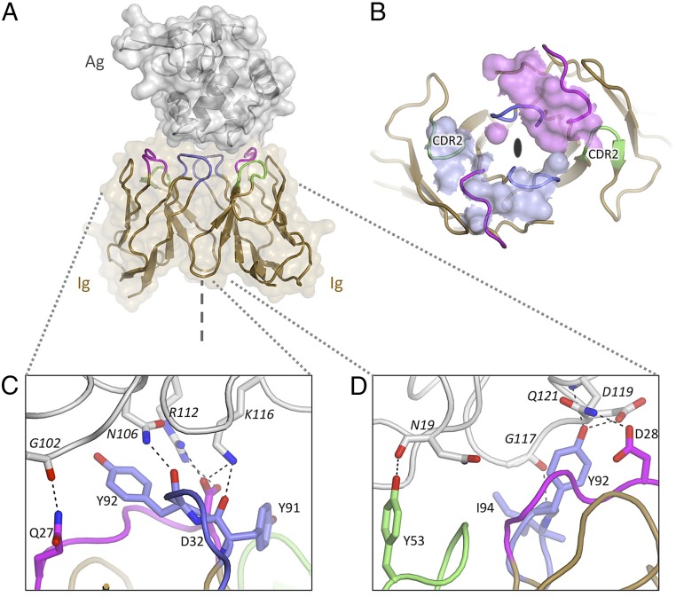
Structure of the Ig12 homodimeric antigen receptor in complex with antigen: Evidence for selective recruitment. (A) Homodimer of Ig domains (tan cartoon with CDR1, CDR2, and CDR3 colored in purple, green, and blue, respectively) bound to a single HEL molecule (gray cartoon and surface). The symmetry axis is indicated (dashed line). (B) HEL contact footprint mapped onto the surface of the Ig12 dimer (viewed down its twofold axis as indicated). The contact surfaces of the two Ig domains are colored in light blue and purple. The CDR2s are indicated and highlight the selective use of otherwise identical surface loops. (C) Antigen-binding site 1 (left-hand side). (D) Antigen-binding site 2 (right-hand side). Hydrogen bonds are highlighted; distinct contact networks are observed for each Ig subunit.

