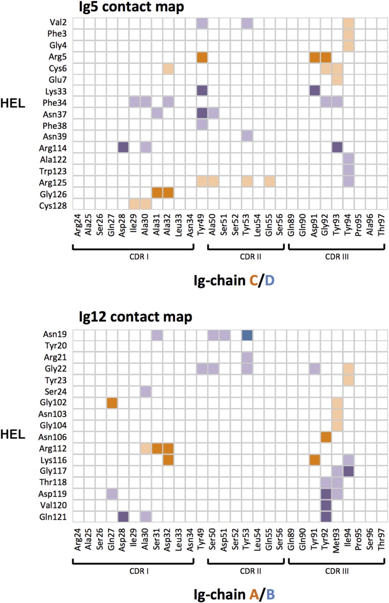
Fig. S5.
Contact map. Surface residues of the two protomers of Ig5 (Top) or Ig12 (Bottom) that contact the surface of HEL. Protomers are distinguished by color (shades of purple or orange, respectively). Light shades indicate hydrophobic interactions. Darker shades indicate hydrogen-bonding interactions or salt bridges. Contact information was derived from analysis output by PDBePISA.

