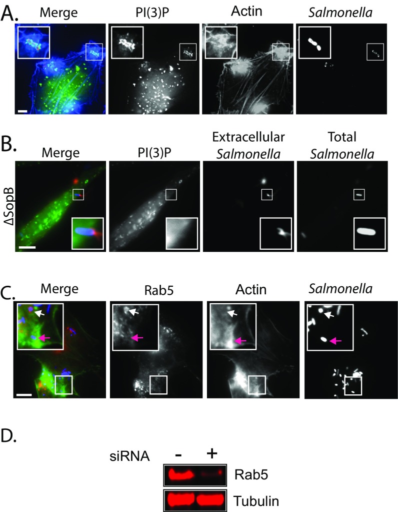
Fig. S3.
(A) Localization of PI(3)P at actin-rich Salmonella membrane ruffles. Cells were transfected with GFP-p40-PX [PI(3)P] and then were infected with Alexa-Fluor 594-labeled wild-type Salmonella (red) and actin stained with Alexa-Fluor 350-conjugated phalloidin (blue). (Scale bar: 5 μm.) Insets 2× magnify the membrane ruffle and invading Salmonella. (B) PI(3)P enrichment at the macropinocytic cup is dependent on SopB. Cells were transfected with GFP-p40Phox (green) and then were infected for 5 min with Alexa-Fluor 350-labeled ΔsopB Salmonella (blue) and extracellular bacteria labeled with anti-Salmonella antibodies (red). (Scale bar: 5 μm.) Insets 3× magnify the macropinocytic cup. (C) Localization of Rab5 during Salmonella infection. Cells were transfected with RFP-Rab5 and then were infected with Alexa-Fluor 350-labeled wild-type Salmonella (blue) and actin stained with Alexa-Fluor 488-conjugated phalloidin (green). Insets 3× magnify infection foci. Purple arrows indicate an invasion site with no Rab5 localization; white arrows indicate Rab5 on Salmonella-containing vacuoles. (Scale bar: 5 μm.) (D) Knockdown efficiency of Rab5. Cells were transfected with nontargeting (−) or Rab5 (+) siRNA before immunoblotting with antibodies against Rab5 and tubulin as a control.

