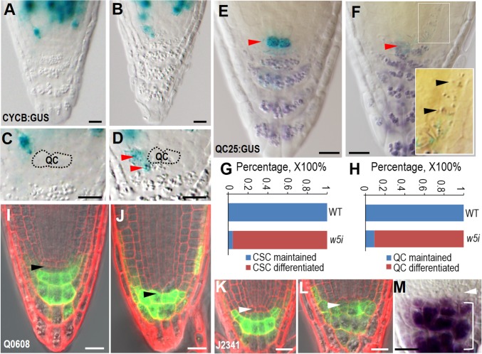
Fig. 2.
Disruption of symplastic signaling in QC causes cell differentiation in the SCN. (A–D) The expression of pCYCB1;1:CYCB1;1-GUS in WT (A and C) and pWOX5:icals3m-expressing roots after 48-h estradiol induction. The QC position is labeled and red arrowheads point to the ectopic cell division marked by the expression of CYCB1;1-GUS in SCN. (E and F) Starch granule accumulation as visualized by Lugol’s staining in the WT (E) and WOX5:icals3m (F) roots after 48-h estradiol induction. Red arrowheads point to the QC position with expression of QC25 marker. Note the starch granules in proximal meristem in zoom-in image in F. (G and H) Quantification of CSC (G) and QC (H) differentiation rate in WT and pWOX5:icals3m (w5i) roots. (I and J) Q0608 expression in WT (I) and pWOX5:icals3m (J) roots after 48-h estradiol induction. Black arrowheads point to the CSC position. (K and L) J2341 expression in WT (K) and pWOX5:icals3m (L) roots after 48-h estradiol induction. White arrowheads point to the QC position. (M) QC (pointed to by the white arrow head) was not differentiated but CSC became differentiated (included in the bracket) in the wox5-1 root. (Scale bars, 20 μm.)

