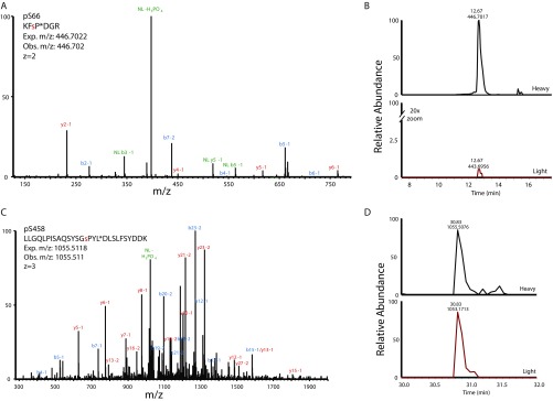
Fig. S6.
Isotopically labeled peptide standards confirm DET1 phosphorylation at Ser66 and Ser458. (A) MS/MS spectrum of isotopically labeled standard peptide representing phosphorylation on the human DET1 protein at Ser66 (pSer66). The red lowercase residue denotes the position of phosphorylation. An asterisk (P*) indicates the isotopically labeled residue. Observed (Obs.) and expected (Exp.) m/z measurements, as well as charge (z) state are shown. Ions from the b' and y' series are indicated on the spectrum in blue and red; ions demonstrating neutral loss (NL) of the phosphate are shown in green. (B) Extracted ion chromatogram (± 10 ppm) for pSer66 showing isotopically labeled standard peptide (Heavy) and the digested analyte peptide (Light). To display coelution more clearly, the y axis of the light peptide was magnified 20×. Retention time (in minutes) and observed m/z for heavy and light peptides are shown above each corresponding peak. (C) MS/MS spectrum of isotopically labeled standard peptide representing phosphorylation on the human DET1 protein at Ser458 (pSer458) with the isotope label incorporated at Leu (L*). (D) Extracted ion chromatogram (± 10 ppm) for pSer458 presented as in B.

