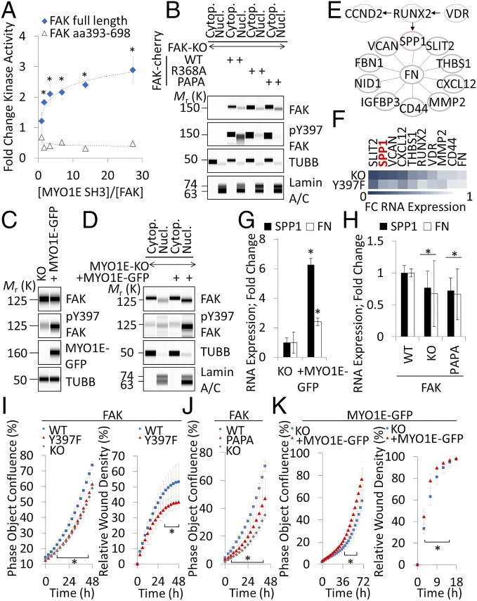
Fig. 4.
MYO1E activates FAK to induce SPP1 expression in fibroblasts. (A) In vitro FAK kinase activity of full-length recombinant FAK versus FAK kinase domain only (aa 393–698) in the presence or absence of 1:2 titrated recombinant MYO1E SH3 (mean ± SEM, n = 4; *P < 0.05 versus no MYO1E SH3). (B) Effects of FAK PRR1 mutations on nuclear (Nucl.) and cytoplasmic (Cytop.) FAK in transiently transfected fibroblasts. (C) Immunodetection of the indicated antigens in stably MYO1E-GFP–reconstituted MYO1E-null mouse embryonic fibroblasts. (D) Nuclear versus cytoplasmic localization of FAK in MYO1E-null (KO) or WT MYO1E-GFP–reconstituted fibroblasts. (E) Network of differentially regulated FN-associated genes. Arrows and lines indicate a functional relationship as determined by STRING. (F) Color-coded ratios of mRNA expression as quantified by RNA sequencing in FAK-null (KO) or Y397F-reconstituted fibroblasts over WT. FC, fold change. RNA expression of the indicated genes in MYO1E-null (KO) or WT MYO1E-GFP–reconstituted fibroblasts (G) or FAK-null fibroblasts transiently reconstituted with WT or PAPA FAK (H) is shown. Gene expression was determined by quantitative PCR (mean ± SD, n ≥ 4; *P < 0.05). (I–K) Phase object confluence or Matrigel invasion of the indicated reconstituted and nonreconstituted KO fibroblasts (mean ± SD, n = 4; *P < 0.05, Student’s t test). Immunoblots are pseudoimages generated by ProteinSimple Compass software.

