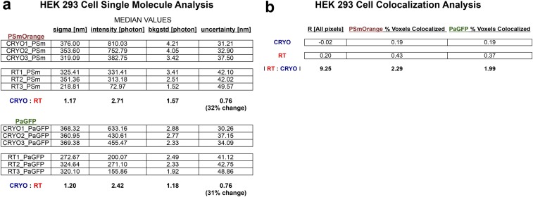
Fig. S3.
Single-molecule and colocalization analyses of PaGFP-Utrophin and PSmOrange-Vimentin in HEK 293 cells at RT and −140 °C. (A) Single-molecule analyses reveal a 2.7–2.4-fold increase in photon yield per molecule that results in a 32–31% decrease in localization uncertainty for PSmOrange and PaGFP at −140 °C vs. at RT. (B) Pearson’s r coefficient (−1 = no pixel overlap, +1 = complete pixel overlap) for regions of interest wherein PaGFP and PSmOrange signals were ≤1 µm from each other within HEK cells. Cells imaged at −140 °C had PaGFP-Utrophin and PSmOrange-Vimentin signals that were 9.25-fold less colocalized. In addition, the percent volume of each signal was between 2–2.3-fold (for PaGFP-Utrophin and PSmOrange-Vimentin, respectively) less colocalized at −140 °C than at RT.

