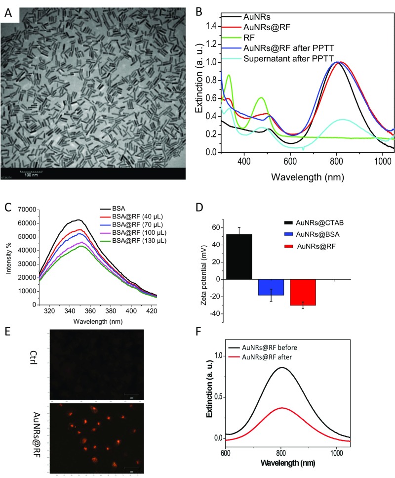
Fig. S1.
Characterization and TU686 cell uptake of RF-conjugated AuNRs@RF. (A) TEM image of conjugated nanorods (AuNRs). (Scale bar, 100 nm.) (B) UV-vis absorption spectra of the as-synthesized AuNRs (black line), BSA and RF conjugated AuNRs@RF (red line), free RF in solution (green line), AuNRs after PPTT (blue), and the supernatant after PPTT (cyan). (C) BSA fluorescent emission quenching of BSA (10−4 M) after adding various amounts of RF (10−4 M) from 0 to 130 μL. (D) Zeta potential of AuNRs with different surface ligands: CTAB, BSA, and BSA + RF. (E) Dark-field microscopic imaging showing TU686 cellular uptake of 2.5 nM AuNRs@RF after 24-h incubation. (Scale bar, 50 μm.) (F) UV-vis absorption spectra of the nanoparticle–media mixture before and after cell culture. For comparison with other studies, 5 nM = 1 OD for small AuNRs (35).

